Your cart is currently empty!
Category: Physical Therapy
-

Shoulder Tendonitis Treatment: Effective Causes and Remedies
Comprehensive Guide to Understanding Shoulder Tendonitis: Identifying Causes, Symptoms, and Proven Treatment Methods
Shoulder tendonitis stands as a prevalent yet often misunderstood ailment that can severely impair the daily routines of countless individuals. This painful condition stems from the inflammation of the tendons encircling the shoulder joint, which is frequently instigated by overuse or certain types of injuries. The resultant inflammation does not merely cause discomfort; it also induces stiffness and limits the range of motion in the shoulder, making even simple actions, such as reaching for a glass or picking up an object, incredibly difficult. For individuals whose jobs or athletic pursuits necessitate extensive use of their shoulders, grasping the intricacies of shoulder tendonitis is essential, as it significantly affects their overall quality of life and their ability to engage in daily activities without experiencing pain.
As we delve deeper into the multifaceted nature of shoulder tendonitis, it becomes increasingly important to acknowledge its widespread prevalence and the various demographics it impacts, which span a wide array of age groups and lifestyles. The shoulder joint’s remarkable flexibility allows for a diverse range of activities, yet this same mobility renders it susceptible to injuries. The rotator cuff, a vital group of muscles and tendons, plays an indispensable role in stabilizing the shoulder while facilitating smooth arm movements. When the tendons within this area become irritated or inflamed, the result can be a debilitating case of shoulder tendonitis. This scenario underscores the importance of early recognition and effective management strategies to mitigate its detrimental effects on daily living.
Key Facts and Insights About Shoulder Tendonitis You Should Be Aware Of
- Shoulder tendonitis is defined by the inflammation of shoulder tendons, which results in pain and limited movement.
- Common contributing factors include repetitive overhead motions, overuse, and poor posture, all of which can lead to chronic symptoms.
- Primary symptoms consist of ongoing pain, stiffness, and weakness in the shoulder area, significantly disrupting daily activities.
- Diagnosis generally involves a comprehensive physical examination, imaging tests, and occasionally shoulder arthroscopy for an exhaustive assessment.
- Non-surgical treatment alternatives, including rest, physical therapy, and anti-inflammatory medications, are often successful in managing shoulder tendonitis.
Identifying the Contributing Factors of Shoulder Tendonitis to Enhance Prevention StrategiesUnderstanding the various factors that contribute to shoulder tendonitis is crucial for effective prevention. One major risk factor is the prevalence of repetitive overhead activities, which many athletes and workers frequently perform. For instance, sports like swimming, tennis, and baseball require extensive shoulder movements that can lead to gradual wear and tear on the tendons. Likewise, occupations that involve repetitive lifting or reaching overhead further heighten the risk of developing this painful condition.
As individuals engage in these activities, they may unknowingly accelerate the wear on their shoulder tendons, leading to inflammation and discomfort over time. Additionally, age-related degeneration plays a significant role; as we grow older, our tendons naturally lose elasticity and strength, making them more prone to injury. Lifestyle factors, such as poor posture and insufficient physical activity, can exacerbate these concerns. Furthermore, certain medical conditions, including diabetes and rheumatoid arthritis, can increase susceptibility to shoulder tendonitis. By recognizing these common risk factors, individuals can take proactive steps to protect their shoulder health and overall well-being.
Identifying the Main Symptoms of Shoulder Tendonitis for Prompt Intervention
The symptoms of shoulder tendinitis can greatly vary in intensity and may manifest differently from person to person. A primary symptom of this condition is persistent pain in the shoulder region, which often worsens with certain movements or activities. This discomfort may feel sharp or dull and can radiate down the arm or even into the neck, complicating daily tasks and diminishing overall comfort.
Individuals may find that their ability to perform everyday activities, such as reaching for objects or lifting items overhead, is severely hindered by their discomfort, impacting both their work and leisure activities. Stiffness in the shoulder joint, especially after periods of inactivity, is another common symptom. This stiffness can further restrict movement, leading to compensatory movements that may worsen the condition. Additionally, swelling around the shoulder joint is frequently observed alongside tendonitis. Recognizing these symptoms early is crucial for seeking timely medical intervention and preventing more serious complications from developing.
Advanced Diagnostic Techniques for Accurate Evaluation of Shoulder Tendonitis
Diagnostic Test Accuracy Cost MRI High High Ultrasound Moderate Moderate Physical Examination Low Low The diagnostic journey for shoulder tendonitis generally begins with a thorough medical history and a physical examination conducted by a qualified healthcare professional. During this assessment, patients will be asked to provide detailed information regarding their symptoms, including when they began and which specific activities might aggravate their discomfort. The physician will conduct a series of tests to evaluate the range of motion and strength of the shoulder joint, which are essential for determining whether tendonitis is the primary issue or if another underlying condition may be contributing to the symptoms.
In some cases, advanced imaging techniques, such as X-rays or MRI scans, may be required to confirm the diagnosis and rule out other potential issues like rotator cuff tears or arthritis. These imaging studies provide vital insights into the condition of the shoulder’s structures, facilitating informed treatment decisions. By understanding the diagnostic process for shoulder tendonitis, individuals can better appreciate the importance of seeking professional evaluation when experiencing persistent shoulder pain.
Exploring Non-Surgical Treatment Options for Effective Relief from Shoulder TendonitisHealthcare professionals frequently recommend non-surgical treatment options as the first line of defense against shoulder tendonitis. One highly effective approach entails a combination of rest and modifications to daily activities. By minimizing or avoiding actions that exacerbate symptoms, individuals grant their inflamed tendons the crucial time needed for healing. In addition to rest, applying ice to the affected area can significantly reduce swelling and alleviate pain, which is particularly important during the early stages of recovery.
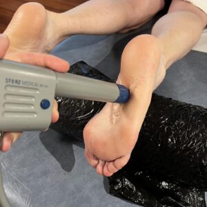
Over-the-counter anti-inflammatory medications can also be beneficial in managing discomfort during this initial phase. Furthermore, engaging in physical therapy plays a pivotal role in the non-surgical treatment landscape. A skilled physical therapist can create a tailored rehabilitation program focused on strengthening the muscles surrounding the shoulder joint while improving flexibility and range of motion. Through targeted exercises and stretching routines, individuals can progressively restore strength and functionality in their shoulders. Additional therapeutic modalities, such as ultrasound therapy or electrical stimulation, may also be utilized to support healing and reduce pain. By exploring these non-surgical options, individuals can actively engage in their recovery while minimizing the need for more invasive treatments.
Examining Surgical Alternatives for Chronic Shoulder Tendonitis: Important Considerations
If non-surgical treatments do not yield relief after several months, or if significant damage to the shoulder structures is present, surgical interventions may become necessary. One commonly performed procedure for addressing shoulder tendonitis is arthroscopy. This minimally invasive technique allows surgeons to visualize and repair damaged tissues within the joint using small incisions and specialized instruments. During an arthroscopic procedure, any inflamed tissue or bone spurs contributing to the patient’s symptoms can be removed, promoting healing and restoring shoulder function.
In some cases, open surgery may be indicated for more extensive repairs or when additional conditions coexist with shoulder tendonitis. This approach requires larger incisions to grant greater access to the shoulder joint for a thorough evaluation and treatment. While surgical procedures can effectively resolve persistent shoulder tendonitis, it is crucial to carefully consider the potential risks and benefits in consultation with a healthcare provider. Additionally, post-operative rehabilitation is vital for recovery, allowing individuals to regain strength and mobility in their shoulders.
Proactive Measures for Preventing Shoulder Tendonitis and Enhancing Joint HealthPreventing shoulder tendonitis is crucial for maintaining optimal shoulder health and avoiding the associated pain of this condition. One highly effective strategy is to engage in regular strength training exercises that specifically target the muscles surrounding the shoulder joint. By increasing strength in these muscles, individuals can provide enhanced support for their shoulders during various physical activities, thereby minimizing the risk of injury.
Moreover, employing proper body mechanics during lifting or overhead movements is essential to reduce strain on the shoulder area. Being aware of one’s physical limits and recognizing when rest is needed can also aid in preventing chronic issues like tendonitis from developing. Ignoring pain signals or overtraining can lead to long-term complications. Additionally, integrating flexibility exercises into a regular fitness regimen can help maintain an optimal range of motion in the shoulders. Stretching before and after physical activities is beneficial in preventing tightness and lowering injury risk. By implementing these proactive preventive measures, individuals can significantly decrease their chances of developing shoulder tendonitis.
Dedication to Recovery and Rehabilitation: Your Path to Overcoming Shoulder Tendonitis
Successfully recovering from shoulder tendonitis requires a strong commitment to a structured rehabilitation program focused on restoring function and alleviating pain. Initially, recovery efforts will emphasize reducing inflammation through rest and ice application, gradually incorporating gentle range-of-motion exercises under professional guidance. As healing progresses, strengthening exercises become increasingly essential for rebuilding muscular support around the shoulder joint.
Throughout the rehabilitation process, it’s vital to remain attentive to the body’s signals and to resist the urge to push too hard too soon. Regular follow-up appointments with healthcare professionals are necessary to monitor progress and adjust the rehabilitation plan as needed. With dedication and adherence to a well-structured recovery program, individuals can expect to regain full functionality in their shoulders while minimizing the risk of future injuries related to tendonitis.
For those seeking further insights into effectively managing shoulder tendonitis, MCR Therapies provides a variety of services that may be especially beneficial. Their treatments focus on alleviating symptoms associated with this condition. Notably, their cupping therapy service is highly regarded for its efficacy in promoting pain relief and muscle healing, making it an excellent option for individuals dealing with shoulder tendonitis.
Provided By: Shoulder Therapy
The Article: Shoulder Tendonitis Causes and Effective Treatments appeared first on https://mcrtherapies.co.uk
The Article Shoulder Tendonitis: Causes and Top Treatment Options appeared first on https://mcrtherapies.com
The Article Shoulder Tendonitis Treatment: Key Causes and Solutions Was Found On https://limitsofstrategy.com
-

Frozen Shoulder: Understanding Causes and Treatment Options
Comprehensive Guide to Frozen Shoulder: Understanding Adhesive Capsulitis and Its Impact
Frozen shoulder, also known in medical terms as adhesive capsulitis, represents a complex and often debilitating condition that profoundly affects daily life and physical activities. This disorder is characterized by persistent stiffness and pain in the shoulder joint, which can greatly restrict an individual’s ability to perform routine tasks and engage in various physical activities. Usually, the onset of frozen shoulder is gradual, and it often develops without a definitive cause. Symptoms may persist for many months or even years, making early awareness of this condition critical. Recognizing the signs early allows for timely interventions that can effectively manage symptoms and significantly enhance quality of life.
In our everyday lives, the significance of shoulder mobility is frequently overlooked until a condition like frozen shoulder emerges. When this condition strikes, it can feel as though our freedom of movement has been severely compromised, leading to frustration and discomfort. Developing a thorough understanding of frozen shoulder is essential; it not only sheds light on the extensive impact this condition can have on our daily existence but also underscores the importance of pursuing appropriate treatment options that aim to restore functionality and alleviate pain.
Understanding the Three Key Phases of Frozen Shoulder: Strategies for Pain Management and Recovery
The progression of frozen shoulder can be effectively divided into three significant phases: the freezing, frozen, and thawing phases. During the initial freezing phase, individuals commonly report increasing pain coupled with a gradual decline in shoulder range of motion, complicating even the most straightforward daily activities. This reduction in mobility can severely disrupt daily life, leading to feelings of helplessness and frustration as individuals find it increasingly challenging to perform tasks that were once effortless.
In the frozen phase that follows, while the pain intensity may stabilize, mobility remains critically restricted, causing ongoing frustration for those affected. The thawing phase marks a pivotal turning point, as individuals begin to regain movement, although this recovery process can be slow and may extend over several months. By thoroughly understanding the distinct phases and progression of frozen shoulder, we can emphasize the importance of early diagnosis and proactive management strategies designed to alleviate its impact and foster recovery.
Key Insights and Important Information About Frozen Shoulder for Enhanced Awareness
- Frozen shoulder, clinically recognized as adhesive capsulitis, is primarily defined by severe pain and stiffness in the shoulder joint, which significantly impedes mobility.
- Common triggers for frozen shoulder include injury, overuse, and various underlying health conditions such as diabetes and thyroid disorders.
- Individuals between the ages of 40 and 60, particularly women, face a higher risk of developing frozen shoulder, often due to hormonal changes and other contributing factors.
- Typical symptoms encompass considerable pain and stiffness, with diagnosis usually confirmed through comprehensive physical assessments and advanced imaging techniques.
- Treatment options for frozen shoulder are diverse, ranging from conservative pain management and physical therapy to more invasive surgical procedures, including manipulation under anesthesia or shoulder arthroscopy.
Analyzing the Underlying Causes of Frozen Shoulder for Better UnderstandingThe exact causes of frozen shoulder remain somewhat elusive; however, this condition is primarily linked to inflammation and thickening of the shoulder capsule, which is vital for joint stability and motion. Various factors, such as injury or extended periods of immobility—often following surgical procedures or due to other medical conditions—can trigger inflammation in this crucial capsule.
This inflammatory response frequently results in the development of scar tissue, which can severely restrict movement and intensify pain. Although initial causes may vary among individuals, the resulting symptoms can be universally debilitating and distressing. In addition to inflammation, numerous underlying medical conditions—including diabetes, thyroid disorders, cardiovascular disease, and Parkinson’s disease—have been associated with a heightened risk of developing frozen shoulder. This highlights the necessity for a multifaceted understanding of the condition’s onset and effective management strategies.
Recognizing the Key Risk Factors Associated with Frozen Shoulder
When examining the risk factors associated with frozen shoulder, it becomes clear that certain demographics are more susceptible than others. Age is a significant factor; those aged 40 to 60 years are considerably more likely to develop this condition. Furthermore, frozen shoulder is particularly prevalent among women, especially during menopause, when hormonal fluctuations can adversely impact the integrity of connective tissues.
Understanding these demographic trends is vital for identifying individuals who could benefit from proactive measures to protect shoulder health. A history of previous shoulder injuries or surgical procedures can also raise the likelihood of developing frozen shoulder. Often, after experiencing surgery or trauma, individuals may unconsciously limit shoulder movement during recovery, thereby increasing the risk of adhesive capsulitis.
Additionally, chronic health conditions, particularly diabetes and autoimmune disorders, can predispose individuals to frozen shoulder due to their effects on inflammation and the body’s tissue repair mechanisms. By recognizing these risk factors, individuals can take proactive steps to mitigate the chances of experiencing this painful condition in the future.
Identifying Symptoms and Effective Diagnostic Approaches for Frozen ShoulderThe symptoms associated with frozen shoulder can often be both perplexing and distressing for those affected. Initially, individuals may experience a dull ache in the shoulder that progressively intensifies over time. This pain can radiate down the arm, disrupting sleep and complicating even the simplest daily activities, such as reaching for items or getting dressed.
As the condition advances, individuals may encounter significant difficulties with basic tasks, leading to heightened frustration and a sense of helplessness. A defining characteristic of frozen shoulder is a pronounced limitation in the range of motion, further complicating everyday life. Diagnosing frozen shoulder typically involves a comprehensive physical examination paired with a detailed medical history.
Healthcare providers will evaluate the range of motion and may conduct specific tests to assess the extent of mobility limitations. Imaging studies, including X-rays or MRIs, may also be utilized to rule out other potential causes of shoulder pain. It is crucial for patients to maintain open communication with their healthcare providers regarding their symptoms and any prior injuries or conditions. A timely and accurate diagnosis is essential for implementing effective treatment strategies aimed at alleviating discomfort and restoring mobility.
Examining a Range of Treatment Options for Effectively Managing Frozen Shoulder
When addressing frozen shoulder, a multifaceted approach is often necessary for effectively managing pain and restoring mobility. Initially, conservative measures such as rest, ice application, and over-the-counter pain medications may be recommended to help control inflammation and alleviate discomfort. These preliminary interventions provide temporary relief while exploring additional rehabilitation options.
Early intervention is critical; addressing symptoms promptly can help prevent further deterioration of the shoulder’s range of motion. As treatment continues, corticosteroid injections may be considered for more severe cases where pain management becomes increasingly challenging. These injections are effective in reducing joint inflammation, providing significant temporary relief from discomfort.
Moreover, physical therapy is frequently recommended as a core component of the treatment strategy. Physical therapy focuses on personalized stretching and strengthening exercises tailored to individual needs, helping to restore mobility while minimizing discomfort. By incorporating various treatment modalities, individuals can work toward achieving optimal outcomes in managing frozen shoulder effectively.
Highlighting the Importance of Physical Therapy and Exercises in Frozen Shoulder RecoveryPhysical therapy plays an essential role in the recovery journey from frozen shoulder, providing targeted interventions designed to improve flexibility and strength in the affected shoulder joint. A qualified physical therapist will conduct a comprehensive assessment of individual needs, developing a customized exercise program specifically tailored to address any limitations. Stretching exercises are particularly vital during this phase, as they work to elongate tight muscles and improve overall range of motion.
In addition to stretching, strengthening exercises that target the surrounding muscles can offer better support for the shoulder joint. Beyond structured exercises, our physical therapist may incorporate therapeutic modalities, such as heat or ultrasound therapy, which can enhance blood circulation and promote healing within the joint. These techniques effectively alleviate pain while preparing muscles for movement.
As therapy progresses, maintaining consistency with a home exercise program becomes essential; commitment to rehabilitation is crucial for achieving lasting results. By actively participating in our recovery journey, we empower ourselves to regain mobility and minimize the impact of frozen shoulder on our daily routines.
Assessing Surgical Options for Frozen Shoulder: Key Factors to Consider
In certain circumstances where conservative treatments fail to provide satisfactory relief or restore functionality, surgical intervention may be necessary. One commonly performed procedure for frozen shoulder is arthroscopic capsular release. During this minimally invasive surgery, the surgeon utilizes specialized instruments to sever the thickened capsule surrounding the shoulder joint, thereby increasing mobility and reducing pain.
While the prospect of surgery may appear daunting, it is typically reserved for cases where all other treatment avenues have been exhausted. Following the surgical procedure, engaging in post-operative rehabilitation is critical to ensure optimal outcomes and regain strength and flexibility in the joint.
Our healthcare team will provide guidance throughout the entire process, assisting us in navigating any challenges that may arise during recovery. While surgical intervention may not be necessary for every individual with frozen shoulder, it remains an important option for those who have not found relief through conservative measures.
Proactive Steps to Prevent Frozen Shoulder: Essential Tips for Optimal Shoulder Health
Preventing frozen shoulder necessitates taking proactive measures to protect shoulder health throughout our lives. One effective strategy for prevention is to maintain an active lifestyle and incorporate regular physical activities that promote shoulder flexibility and strength. Simple exercises, such as arm circles or gentle stretches, can help maintain joint mobility and avert stiffness.
Furthermore, for individuals who have previously experienced an injury or undergone shoulder surgery, adhering to recommended rehabilitation exercises is crucial to ensure a complete recovery. Being vigilant about underlying health issues that may increase the risk of developing frozen shoulder is equally important. For those managing diabetes or other chronic conditions, effectively controlling these health issues can significantly lower the risk of complications related to adhesive capsulitis.
By prioritizing overall health through routine check-ups and lifestyle adjustments, we can take substantial steps to prevent frozen shoulder from affecting our daily lives. Ultimately, awareness and proactive strategies are critical components in safeguarding our shoulder health as we age.
If you’re interested in exploring alternative treatment options for frozen shoulder, consider investigating cupping therapy. This alternative medical technique involves placing cups on the skin to create suction, potentially alleviating pain and reducing inflammation. To learn more about how cupping therapy may benefit individuals with conditions like frozen shoulder, visit MCR Therapies’ cupping therapy page. This resource provides comprehensive insights about the procedure and its potential benefits.
Presented By: Frozen Shoulder Therapy
The Article: Frozen Shoulder: Causes and Effective Treatment Options appeared first on https://mcrtherapies.co.uk
The Article Frozen Shoulder: Understanding Causes and Treatment Options appeared first on https://mcrtherapies.com
The Article Frozen Shoulder Causes and Treatment Options Explained Was Found On https://limitsofstrategy.com
-

Instrument-Assisted Soft Tissue Mobilisation Aids Recovery
Enhancing Recovery with Instrument-Assisted Soft Tissue Mobilisation
Healthcare practitioners employ a manual therapy technique known as Instrument-Assisted Soft Tissue Mobilisation (IASTM) to effectively treat soft tissue injuries and musculoskeletal issues. This method utilises specially designed instruments, typically crafted from stainless steel, to pinpoint and rectify areas of compromised soft tissue function. The devices apply targeted pressure and friction to the affected areas, promoting the dissolution of scar tissue, adhesions, and restrictions within the fascia.
IASTM, or Instrument-Assisted Soft Tissue Mobilisation, is a minimally invasive technique used to treat various conditions, including tendinitis, muscle strains, ligament sprains, and myofascial pain syndrome. Additionally, it has shown effectiveness in addressing chronic conditions like plantar fasciitis, carpal tunnel syndrome, and tennis elbow. The primary objectives of IASTM are to improve soft tissue function, reduce discomfort, and restore normal movement patterns.
The main objective of IASTM is to offer patients lasting relief from symptoms by targeting the underlying causes of soft tissue dysfunction. IASTM is applicable to a wide range of patients and is frequently incorporated into holistic treatment plans in conjunction with various therapeutic methods. Although IASTM has become increasingly popular in recent years, it is important to note that the success of this technique can be influenced by individual conditions and patient characteristics.
Main Points
- IASTM is a hands-on therapeutic approach designed to address soft tissue injuries and musculoskeletal disorders. The process entails the mobilisation and manipulation of soft tissue through the use of specially crafted tools.
- IASTM offers several advantages for recovery, such as enhanced blood circulation, decreased pain and inflammation, greater range of motion, and faster healing of soft tissue injuries.
- IASTM facilitates the healing of soft tissues by effectively breaking down scar tissue, stimulating collagen production, and enhancing the alignment and function of tissues.
- IASTM is essential in rehabilitation and injury prevention, effectively targeting soft tissue restrictions, enhancing tissue quality, and improving overall movement patterns.
- Techniques and tools frequently utilised in IASTM encompass scraping, cross-friction massage, and instruments like Graston and HawkGrips.
- When utilising IASTM, it is essential to take precautions such as steering clear of regions with open wounds, infections, or acute inflammation. Additionally, ensuring proper training and technique is crucial to avoid any potential tissue damage.
- Incorporating IASTM into a holistic recovery strategy entails blending it with additional techniques like exercise, stretching, and manual therapy to tackle the root causes of soft tissue dysfunction and foster sustainable recovery.
The Benefits of IASTM for RecoveryAnalysing Scar Tissue and Adhesions
IASTM provides a notable benefit in the healing process of soft tissue injuries and musculoskeletal issues by effectively breaking down scar tissue and adhesions that may form in the soft tissues after an injury. Adhesions may limit mobility and lead to discomfort, rigidity, and diminished functionality. Utilising IASTM to address these adhesions enables healthcare professionals to assist patients in restoring normal movement patterns and alleviating pain.
Enhancing Circulation and Facilitating Recovery
One more advantage of IASTM is its capacity to enhance blood circulation to the targeted region. Enhanced blood circulation facilitates the healing process by supplying vital nutrients and oxygen to the tissues while effectively eliminating waste products and toxins. This can aid in diminishing inflammation, swelling, and discomfort, while also promoting the body’s inherent healing mechanisms.
Enhancing Collagen Production and Promoting Tissue Repair
Moreover, IASTM can promote collagen production, a crucial factor in the repair and regeneration of damaged tissues. This may result in a quicker and more efficient recovery from soft tissue injuries and musculoskeletal conditions.
How IASTM Enhances Soft Tissue Healing
IASTM promotes the healing of soft tissues by activating the body’s natural healing processes and reducing the barriers that impede recovery. Soft tissue injuries may result in the development of scar tissue and adhesions as a consequence of the body’s natural healing mechanisms. While these tissues are essential in the initial phases of wound healing, improper positioning or restriction of movement can lead to complications.
IASTM aids in the realignment of scar tissue and the breakdown of adhesions, fostering improved tissue healing and reduced discomfort. IASTM, or Instrument-Assisted Soft Tissue Mobilisation, effectively breaks down scar tissue and adhesions while improving the flexibility and suppleness of soft tissues. This approach can promote the recovery of normal movement patterns and reduce the chances of re-injury.
Restoring flexibility, range of motion, and strength to the soft tissues is crucial for a complete recovery, and IASTM offers effective assistance in this process. Furthermore, IASTM can effectively reduce inflammation in the affected area. Inflammation is a natural response to injury; however, an excessive or prolonged inflammatory reaction can impede the healing process.
Instrument-Assisted Soft Tissue Mobilisation (IASTM) is a therapeutic approach designed to promote tissue healing through enhanced blood circulation and reduced inflammation. Enhancing blood circulation, IASTM guarantees that the impacted tissues obtain the essential nutrients and oxygen required for a swift recovery. This knowledge equips you, the healthcare expert, to make well-informed decisions regarding the treatment of soft tissue injuries.
The Role of IASTM in Rehabilitation and Injury PreventionIASTM, or Instrument-Assisted Soft Tissue Mobilisation, plays a crucial role in the rehabilitation process. This method effectively restores normal function to the soft tissues, addressing the source of pain and impaired function. Rest and traditional rehabilitation methods frequently fall short in fully addressing soft tissue injuries. IASTM provides a targeted approach for addressing scar tissue, adhesions, and fascial restrictions that may hinder a patient’s recovery.
Additionally, IASTM serves as a proactive approach to reduce the risk of future injuries. Healthcare providers play a crucial role in helping patients maintain optimal musculoskeletal health and function by proactively addressing soft tissue dysfunction before it develops into a more serious injury. This can yield beneficial outcomes, especially for individuals and athletes. Individuals engaged in repetitive or high-impact activities may face the risk of soft tissue injury.
Incorporating Instrument-Assisted Soft Tissue Mobilisation (IASTM) into a well-rounded rehabilitation program can greatly enhance patient relief, facilitating a faster and more complete recovery from injuries. Instrument-Assisted Soft Tissue Mobilisation (IASTM) is an effective method for addressing soft tissue dysfunction, focussing on both the symptoms and underlying causes. This method allows individuals to regain their natural movement patterns, reduce discomfort, and improve their overall quality of life, offering both comfort and peace of mind.
Common Techniques and Tools Used in IASTM
IASTM employs a range of techniques and tools to effectively tackle soft tissue injuries and musculoskeletal issues. A commonly utilised technique is known as “scraping” or “cross-friction massage.” This technique employs a meticulously designed instrument to apply pressure and massage the affected area, effectively breaking down scar tissue and adhesions. The device oscillates accurately across the skin to address the impaired soft tissues.
A frequently utilised technique in IASTM is known as “stroking” or “flushing.” This technique involves utilising the device to apply gentle pressure through broad, sweeping movements across the entire affected muscle or tendon. This method improves blood flow to the targeted area, reduces inflammation, promotes relaxation, and eases discomfort. IASTM employs instruments primarily made from stainless steel or other durable materials, allowing for precise management and manipulation of soft tissues.
These instruments come in a variety of shapes and sizes to suit different body areas and types of injuries. Healthcare professionals receive training to guarantee the safe and efficient use of these instruments, enabling them to deliver precise treatment to their patients.
Precautions and Considerations for IASTM
Contraindications and Risk Factors
Individuals with specific medical conditions or risk factors might not be ideal candidates for IASTM. For example, people with bleeding disorders or those on blood-thinning medications might face a higher risk of complications from IASTM.
Customised Care and Engagement
Healthcare professionals are required to evaluate the unique needs of each patient and customise their treatment plans accordingly. It is crucial to closely monitor each patient’s progress with IASTM, as responses can vary significantly, and adjustments may be necessary to optimise outcomes. Furthermore, patients are encouraged to express any discomfort or concerns they experience during or following IASTM treatment.
Appropriate Training and Certification
Proper training and certification in IASTM are essential for healthcare professionals to guarantee safe and effective patient treatment. This involves comprehending the indications and contraindications of IASTM and effectively utilising the tools to attain the best possible outcomes.
Integrating IASTM into a Comprehensive Recovery PlanIntegrating Instrument-Assisted Soft Tissue Mobilisation (IASTM) into a comprehensive rehabilitation plan can significantly improve treatment results for patients. IASTM serves effectively as a standalone treatment or can be integrated with various manual therapy techniques, such as massage therapy, joint mobilisation, and therapeutic exercise. This thorough strategy can successfully address both the symptoms and underlying factors of soft tissue dysfunction, improving pain management, functionality, and overall quality of life.
Furthermore, integrating Instrument Assisted Soft Tissue Mobilisation (IASTM) into a comprehensive rehabilitation program could help patients achieve lasting results by targeting both immediate symptoms and the underlying factors that may be contributing to their condition. IASTM offers enduring relief from symptoms by improving the functionality of soft tissues and promoting repair at the cellular level. In conclusion, Instrument Assisted Soft Tissue Mobilisation (IASTM) serves as an effective approach for tackling soft tissue injuries and musculoskeletal issues.
Instrument-Assisted Soft Tissue Mobilisation (IASTM) is an effective technique that improves patient functionality, reduces pain, and accelerates healing by addressing scar tissue, adhesions, and restrictions in the fascia. When integrated into a holistic rehabilitation strategy, IASTM may provide lasting benefits for individuals seeking relief from their symptoms.
FAQs
What is Instrument-Assisted Soft Tissue Mobilisation (IASTM)?
Instrument-Assisted Soft Tissue Mobilisation (IASTM) is a manual treatment technique used by healthcare practitioners to detect and treat soft tissue disorders. Specially developed devices are used to mobilise and manipulate soft tissue in order to promote healing and alleviate discomfort.
How does Instrument-Assisted Soft Tissue Mobilisation work?
IASTM reduces scar tissue and fascial limitations in the afflicted region. IASTM tools aid in the detection and treatment of soft tissue dysfunction by increasing blood flow, decreasing inflammation, and facilitating tissue repair.
What problems may be addressed with Instrument Assisted Soft Tissue Mobilisation?
IASTM is effective in treating musculoskeletal problems such as tendinopathies, muscular strains, ligament sprains, fascial limitations, and scar tissue adhesions. It is widely utilised in sports medicine and physical therapy settings.
Is Instrument-Assisted Soft Tissue Mobilisation Painful?
IASTM may produce pain during therapy, particularly in regions of tissue dysfunction. However, the pain is generally minor and transient. Patients may feel bruising and pain following the therapy, although this usually goes away within a few days.
Who can conduct Instrument-Assisted Soft Tissue Mobilisation?
IASTM should only be conducted by qualified healthcare experts, such as physical therapists, chiropractors, or sports medicine specialists. Proper training and certification are required for safe and successful treatment.
Brought To You By:
References
- Instrument-assisted soft tissue mobilization (IASTM) https://www.physio-pedia.com/Instrument_Assisted_Soft_Tissue_Mobilization
- Therapeutic effectiveness of instrument-assisted soft tissue mobilization https://www.ncbi.nlm.nih.gov/pmc/articles/PMC5331993/
The Article: Instrument-Assisted Soft Tissue Mobilisation Improves Recovery appeared first on https://mcrtherapies.co.uk
The Article Instrument-Assisted Soft Tissue Mobilisation Aids Recovery appeared first on https://mcrtherapies.com
The Article Instrument-Assisted Soft Tissue Mobilisation Aids Recovery Was Found On https://limitsofstrategy.com
-

Fascial Distortion Model: Effective FDM Treatment
Fascial Distortion Model: A Proven Approach to Treatment
The Fascial Distortion Model (FDM) is a diagnostic and treatment approach that emphasises identifying and rectifying distortions in the body’s connective tissue, called fascia. Fascia is a complex network of collagen and elastin fibres that supports muscles, organs, and structures throughout the body. Fascia distortion can lead to various symptoms, including pain, limited movement, and dysfunction in the affected area.
The FDM was created by American osteopathic physician Stephen Typaldos, D.O., in the 1990s. Dr. Typaldos noted that conventional medical approaches did not adequately address many musculoskeletal injuries and conditions, prompting him to develop a groundbreaking model for understanding and tackling these issues. The FDM categorises fascial distortions into six distinct types, each displaying unique symptoms and patterns of dysfunction.
The categories encompass Triggerband, Continuum Distortion, Cylinder Distortion, Herniated Triggerpoint, Folding Distortion, and Tectonic Fixation. The FDM distinguishes itself by focusing on the identification and treatment of specific fascial distortions through hands-on techniques rather than relying solely on imaging or other diagnostic approaches. Skilled practitioners in the FDM utilise palpation and observation techniques to identify areas of distortion within the fascia, subsequently applying focused manual therapies to address these issues effectively.
The FDM targets the underlying fascial distortions to alleviate pain, enhance function, and restore mobility in the affected body areas.
Summary
- The Fascial Distortion Model (FDM) serves as a comprehensive diagnostic and treatment framework focused on identifying and managing specific types of fascial distortions within the body.
- Fascial distortions can be identified through a comprehensive physical examination and a detailed patient history assessment—these issues frequently present as discomfort, restricted mobility, or additional symptoms.
- Treatment methods for fascial distortions can include manual therapy techniques such as manipulation, myofascial release, and exercise and movement therapies.
- The Fascial Distortion Model presents numerous benefits, including improved pain relief, increased range of motion, and faster injury recovery.
- Case studies and success stories demonstrate the effectiveness of FDM in tackling various musculoskeletal conditions and injuries.
- Integrating the Fascial Distortion Model with supplementary treatment approaches, such as physiotherapy and chiropractic care, provides a comprehensive strategy for managing patient care.
- Finding a qualified expert for Fascial Distortion Model treatment is crucial, necessitating a search for healthcare professionals with specialised training and FDM experience.
Identifying Fascial Distortions in the BodyAssessment Techniques
Experts skilled in FDM employ palpation and observation methods to identify specific types of fascial distortions guided by unique symptoms and patterns of dysfunction. For example, a Trigger and distortion can present as a taut, rope-like band of tissue that causes sharp, shooting pain with movement. Conversely, cylinder distortion can manifest as a swollen, sausage-shaped area of tissue that restricts movement and leads to aching or throbbing discomfort.
Assessment of Mobility and Diagnostic Imaging
In addition to palpation and observation, practitioners may employ movement assessments to identify areas of fascial distortion. By meticulously observing patients’ movements and how they perform tasks, practitioners can pinpoint areas of restricted mobility or dysfunctional movement patterns that may indicate underlying fascial distortions. Imaging studies, such as MRI or ultrasound, may be employed to confirm the presence of fascial distortions in specific cases; however, these methods are not always necessary for diagnosis.
Attention and Rehabilitation
Upon recognising fascial distortions, practitioners can develop a customised treatment plan that specifically targets these issues through focused manual therapies. These therapies may include techniques such as trigger and release, cylinder distortion reduction, or tectonic fixation reduction, all designed to precisely address each type of fascial distortion. By addressing these distortions, practitioners aim to relieve pain, improve function, and restore mobility in the affected area.
Treatment Techniques for Fascial Distortions
The treatment techniques utilised in the Fascial Distortion Model are based on precise manual therapies designed to address each type of fascial distortion specifically. These techniques encompass practical methods such as soft tissue manipulation, stretching, and mobilisation to relieve tension, reduce swelling, and restore normal function in the affected area. For example, Triggerband release techniques may involve applying pressure and stretching to the specific area to relieve tight tissue bands and reduce discomfort. Conversely, strategies for lowering cylinder distortion may employ gentle compression and mobilisation to alleviate swelling and improve mobility.
In addition to these specialised techniques, experts trained in FDM may integrate supplementary therapies such as ice or heat application, electrical stimulation, or therapeutic exercise to amplify the advantages of manual treatment and promote healing in the affected area. These complementary therapies are employed with manual techniques to address pain, inflammation, and dysfunction associated with fascial distortions. Treatment techniques in the FDM are tailored to each patient, considering their type of fascial distortion, individual symptoms, and patterns of dysfunction.
This customised approach allows professionals to effectively address the underlying pain and dysfunction issues, leading to enhanced patient results.
Benefits of the Fascial Distortion ModelThe Fascial Distortion Model offers various benefits for those seeking relief from musculoskeletal pain and dysfunction. The FDM provides a notable benefit by focusing on the identification and treatment of specific fascial distortions that conventional medical approaches may overlook or misdiagnose. By tackling these core issues, the FDM aims to provide more effective relief from pain and dysfunction than traditional treatments, which frequently concentrate only on symptoms rather than addressing the root cause.
The FDM offers a significant benefit by emphasising practical manual therapies, which can provide immediate pain relief and improve functionality in the affected area. Many patients experience significant pain relief and improved mobility after just one or two sessions with a practitioner experienced in the FDM approach. This prompt response to treatment can be especially beneficial for those who have experienced chronic pain or limited mobility for an extended period.
FDM offers a customised treatment strategy that considers each patient’s unique symptoms and dysfunction patterns. By tailoring treatment approaches to address specific types of fascial distortions in each individual, practitioners proficient in FDM can provide more targeted and effective care that addresses the underlying causes of pain and dysfunction.
Case Studies and Success Stories
Many case studies and success stories illustrate the efficacy of the Fascial Distortion Model in relieving musculoskeletal pain and dysfunction. A case study published in the Journal of Bodywork and Movement Therapies presented a patient experiencing chronic low back pain who achieved significant relief after just two sessions of FDM treatment. The patient had pursued various conventional therapies without attaining the expected outcomes. Following FDM treatment targeting specific fascial distortions in the lower back, they reported a notable reduction in pain and improved mobility.
A case study published in the International Journal of Osteopathic Medicine examined a patient experiencing shoulder pain and restricted range of motion. A notable enhancement was observed after FDM treatment, which focused on correcting a cylinder distortion in the shoulder girdle. The patient reported significant pain relief and improved functionality after just one session of FDM treatment, underscoring the rapid effectiveness of the targeted manual therapies utilised in FDM. This compilation of case studies and success stories highlights the potential for fast relief from musculoskeletal pain and dysfunction through targeted manual therapies based on the Fascial Distortion Model.
People dealing with chronic pain or limited mobility can find significant relief and improved function after receiving FDM treatment from a qualified professional.
Integrating the Fascial Distortion Model with Other Treatment Modalities
Collaborative Assistance
Experts trained in the FDM often collaborate with other healthcare professionals, such as physiotherapists, chiropractors, and osteopaths, to develop a customised treatment plan that thoroughly addresses a patient’s condition.
Enhanced Treatment Outcomes
Combining the FDM with other evidence-based therapies, such as therapeutic exercise, chiropractic adjustments, or acupuncture, enables patients to achieve enhanced treatment outcomes and ongoing healing and recovery. This detailed approach can improve outcomes by addressing the complex needs of musculoskeletal pain and dysfunction individuals.
Tailored Treatment Solutions
Integrating FDM with supplementary modalities provides patients with an expanded range of treatment options, enabling tailored solutions to address their needs and conditions. Individuals facing intricate musculoskeletal challenges may benefit from integrating FDM treatment with physiotherapy to address strength and mobility deficits or combining it with acupuncture to reduce pain and inflammation.
Finding a Qualified Practitioner for Fascial Distortion Model TreatmentIndividuals looking for alleviation from musculoskeletal pain and dysfunction should seek out a qualified practitioner in the Fascial Distortion Model treatment. Individuals with advanced training in FDM have the knowledge and skills to identify specific fascial distortions and apply targeted manual therapies to address these issues efficiently. When seeking a qualified practitioner for FDM treatment, patients should look for healthcare providers who have completed advanced training in FDM from reputable organisations, such as the American Fascial Distortion Model Association (AFDMA) or other recognised training institutions.
These courses provide practitioners with a comprehensive understanding of FDM principles, effective assessment techniques, and customised treatment protocols designed for each type of fascial distortion. Patients can receive referrals from their primary care physician or other healthcare professionals to locate qualified practitioners offering FDM treatment in their area. Additionally, online directories or industry associations may provide listings of practitioners who have completed advanced training in the FDM and are qualified to offer this specialised form of care.
Choosing a qualified expert for FDM treatment enables patients to receive customised manual therapies designed to address their specific fascial distortions, promoting rapid alleviation of musculoskeletal pain and dysfunction. This tailored care method can lead to more effective results for those seeking relief from chronic pain or limited mobility.
FAQs
What does the fascial distortion model (FDM) treatment entail?
The fascial distortion model (FDM) is a manual therapy approach designed to identify and relieve musculoskeletal pain and dysfunction by addressing distortions in the body’s connective tissue, called fascia.
What is the mechanism behind FDM treatment?
FDM treatment employs precise manual techniques to successfully release and realign distorted fascial tissues, relieve pain, improve mobility, and restore
Which conditions can FDM treatment assist with?
FDM treatment is commonly employed to address a range of musculoskeletal concerns, including back pain, neck pain, joint pain, sports injuries, and repetitive strain injuries.
Is FDM treatment appropriate for all individuals?
FDM treatment may not be suitable for everyone. It is essential to consult with a qualified healthcare expert to evaluate its appropriateness for your specific condition and medical history.
What potential side effects might arise from FDM treatment?
FDM treatment is generally considered safe; however, some individuals may experience temporary soreness or discomfort following a session. It is crucial to address any concerns with a qualified practitioner before undergoing FDM treatment.
What is the usual number of FDM treatment sessions required?
The number of FDM treatment sessions needed can vary depending on the individual and the details of their condition. Some individuals may observe significant improvement after just a few sessions, while others may require ongoing treatment to achieve their desired outcomes.
Brought To You By:
The Article: Fascial Distortion Model: Effective FDM Treatment appeared first on https://mcrtherapies.co.uk
The Article Fascial Distortion Model: Effective FDM Treatment appeared first on https://mcrtherapies.com
The Article Fascial Distortion Model: Effective FDM Treatment Was Found On https://limitsofstrategy.com
-

Dry Needling & Manual Therapy
Enhancing Manual Therapy with Dry Needling
Physical therapists, chiropractors, and other medical professionals use dry needling as an effective therapeutic technique for addressing musculoskeletal pain and movement disorders. Tiny needles are strategically inserted into targeted pressure points on the body, relieving stress and pain. Dry needling is grounded in Western medicine and neurophysiology, distinguishing it from acupuncture, rooted in traditional Chinese medicine.
The primary goal of dry needling is to locate and address trigger points and hyperirritable areas in skeletal muscle that may result in noticeable nodules or tense bands. These trigger points can often cause localised and referred pain, restricted movement, and muscle weakness. Nevertheless, dry needling provides a glimmer of hope and positivity by alleviating pain and enhancing functional ability by releasing these trigger points.
When it comes to comprehensive treatment plans that include physical therapy, exercise, and other forms of treatment, dry needling is a safe and effective procedure that plays a crucial role. This task should only be carried out by highly trained and certified healthcare professionals who have undergone specialised training to ensure the safety and well-being of the patient. You can be confident that patients will receive clear and thorough information regarding the potential advantages and risks of the procedure before undergoing therapy.
This approach is effective in addressing musculoskeletal pain and can be combined with other manual therapy interventions.
Main Points
- Dry needling is a highly effective method for alleviating pain and enhancing functionality. It entails the precise insertion of thin needles into specific muscle trigger points.
- The science behind dry needling revolves around stimulating the nervous system and encouraging the release of natural pain relievers to alleviate muscle tension and enhance blood circulation.
- Combining dry needling with manual therapy can significantly improve treatment results by effectively targeting the soft tissue pain associated with musculoskeletal pain.
- Utilising dry needling techniques, muscle trigger points are effectively targeted to alleviate pain and enhance the range of motion. This makes it an invaluable resource for effectively managing muscle pain.
- Musculoskeletal pain can be effectively addressed by incorporating dry needling alongside joint mobilisation techniques. This approach targets the affected tissue and joint structures, providing a comprehensive treatment solution.
The Science Behind Dry Needling: How it Works to Relieve painLocal Twitch Response and Muscle Relaxation
A needle inserted into a trigger point can elicit a local twitch reaction, causing the muscles surrounding the needle to contract reflexively. This twitch reaction is thought to help relieve muscle tension and break the cycle of dysfunction and discomfort.
Exploring the Benefits of Pain Relief and Neurophysiological Effects
When the needle is inserted, it can stimulate the body’s natural painkillers, known as endorphins. This leads to quick pain relief and a decrease in muscle soreness. In addition, dry needling can potentially impact the nervous system by altering sensory nerve activity and reducing the transmission of pain signals to the brain. This can help desensitise the nervous system and minimise the sensation of pain.
Understanding the Process of Local Blood Flow and Tissue Healing
Dry needling induces microtrauma at the insertion site, leading to enhanced local blood flow and improved tissue repair. Enhancing the oxygen and nutrient levels in the area while eliminating metabolic waste products can have a positive impact. Understanding the science behind dry needling allows for a deeper appreciation of its impact on the body, such as reducing pain and improving overall function.
Integrating Dry Needling with Manual Therapy: Enhancing Treatment Outcomes
The effectiveness of treating musculoskeletal pain and dysfunction can be enhanced by incorporating manual therapy techniques like joint mobilisation, soft tissue mobilisation, and stretching dry needling. Manual therapy aims to restore the natural movement of joints and soft tissues, while dry needling relieves trigger points and muscle tension. By combining these approaches, healthcare practitioners can effectively tackle both the mechanical and neuromuscular aspects of pain and movement limitations.
Suppose a patient is experiencing symptoms of shoulder pain and limited mobility due to a rotator cuff injury. In that case, a physical therapist may use manual techniques to improve joint mobility. Additionally, they may use dry needling to target trigger points in the muscles and provide relief. By combining both methods, you can experience enhanced pain relief, increased range of motion, and improved functional activities. In addition, the combination of dry needling and manual therapy can help prolong the benefits of treatment by addressing both the root cause of musculoskeletal dysfunction and the resulting pain.
Healthcare providers should understand manual therapy and dry-needling techniques to integrate them into treatment programs successfully. Just as crucial is educating patients, as it plays a vital role in helping individuals grasp these therapies’ purpose and anticipated outcomes. By integrating these treatments, healthcare practitioners can provide comprehensive therapy to patients experiencing musculoskeletal discomfort.
Targeting Trigger Points: Using Dry Needling for Muscle Pain Relief
Utilising dry needling techniques, muscle trigger points can be effectively targeted to alleviate muscle discomfort and dysfunction. Palpable nodules or taut bands often accompany hyperirritable areas in skeletal muscle. These websites may lead to localised and transferred discomfort, limited range of motion, and muscle atrophy.
Healthcare providers are crucial in ensuring a safe and effective dry-needling procedure. By strategically inserting tiny needles into specific trigger points, a local twitch response is triggered, releasing muscle tension and relieving patients. For instance, if a patient is experiencing low back pain that worsens with prolonged sitting, a physical therapist’s professional knowledge can help identify trigger points in the paraspinal muscles that may be causing the discomfort. A skilled practitioner can effectively relieve pain and tension by utilising dry-needling techniques in these specific areas.
In addition, utilising dry needling to address trigger points can improve muscle flexibility and function, allowing individuals to experience greater mobility and perform daily tasks with less discomfort. An in-depth assessment by healthcare professionals is essential to pinpoint trigger points and determine if dry needling is a suitable treatment option. It is important to provide patients with clear information about what they can expect during their dry needling therapy, including any possible side effects or warnings.
Healthcare providers have the expertise to address muscular discomfort and dysfunction by focusing on trigger points through dry needling, a therapy that has been proven effective.
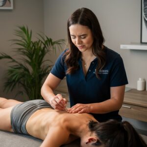
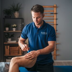
Combining Dry Needling with Joint Mobilization: Addressing Musculoskeletal PainCombining joint mobilisation procedures with dry needling can effectively address musculoskeletal pain and movement limitations by targeting trigger points within muscles. Joint mobilisation involves applying precise pressures to a joint to restore its natural range of motion and provide relief from pain. By incorporating physical needling into joint mobilisation therapies, healthcare professionals can efficiently address both the muscular and articular components of musculoskeletal disorders.
For example, if a patient is suffering from knee pain due to patellofemoral dysfunction, a physical therapist can use joint mobilisation techniques to improve the movement of the patella. In addition, they can use dry needling to relieve trigger points in the quadriceps muscles. This comprehensive approach can alleviate discomfort and improve functional activities like walking, climbing stairs, and squatting. By addressing the joint and muscular components of dysfunction, healthcare practitioners can enhance patient care and provide a more comprehensive approach.
Healthcare providers should thoroughly understand joint mobilisation and dry-needling procedures to effectively integrate them into treatment programs. Equally crucial is educating patients, encouraging them to care for themselves and prioritising their long-term well-being. It assists individuals in comprehending the rationale behind these interventions and the anticipated treatment results. By incorporating these methodologies, medical professionals can provide a more comprehensive approach to treating individuals with musculoskeletal discomfort. This approach emphasises a dedication to their patients’ long-term health and well-being.
Addressing Neuropathic Pain: The Role of Dry Needling in Nerve Pain Management
Gaining Insight into Neuropathic Pain
Neuropathic pain is a complex condition that occurs when the neurological system is damaged or impaired. These symptoms can cause shooting or burning sensations, numbness, or tingling. Neuropathic pain trigger points can be located either along nerve pathways or in muscles supplied by the affected nerves.
Dealing with Sciatica through Dry Needling
For example, people with sciatica may suffer from neuropathic pain caused by compression or irritation of the sciatic nerve. By strategically identifying and treating specific trigger points along the sciatic nerve pathway, skilled healthcare practitioners can effectively alleviate distressing symptoms like leg pain and numbness. This method can alleviate neuropathic pain and enhance functionality by relieving tension in nerve trigger points.
Maximising the Benefits of Dry Needling for Nerve Pain Relief
Healthcare providers need to have a thorough understanding of nerve anatomy and neuropathic pain syndromes to effectively use dry needling to manage nerve pain. They should also provide patients with comprehensive information about the treatment process and any potential risks or precautions that may be associated with dry needling on nerve-related trigger points. Healthcare providers have the expertise to effectively address nerve-related symptoms by using dry needling to target neuropathic pain.
Patient Education and Safety: Exploring the Benefits and Risks of Dry Needling in Manual TherapyPatient education is essential when incorporating dry needling into manual therapy treatments. It is crucial for individuals undergoing dry needling to have a thorough understanding of the therapy’s potential benefits and risks. Healthcare providers should prioritise dedicating ample time to explaining the intricacies of dry needling, offering a thorough explanation of the treatment procedure, and openly discussing any potential adverse effects, such as temporary discomfort or discolouration at the needle insertion sites.
In addition, educating patients about the safety precautions related to dry needling is essential. It is important to avoid strenuous physical activity immediately after the procedure and promptly notify the healthcare provider of any unusual or prolonged symptoms. Patients must understand that dry needling can be a valuable treatment option for musculoskeletal discomfort, but it may not suit everyone or every condition. Healthcare providers can enhance patient comfort during sessions by improving patient education on dry needling and empowering them to make well-informed decisions regarding their treatment options. This, in turn, can effectively reduce anxiety and significantly improve the overall treatment experience.
In addition, it is crucial to foster transparent and open communication between patients and healthcare professionals to address any concerns or questions related to dry needling effectively. Patient education is crucial for ensuring safety and achieving positive outcomes when incorporating dry needling into manual therapy procedures. In manual treatment strategies, dry needling is a highly advantageous method for addressing musculoskeletal pain and movement limitations.
Healthcare practitioners can enhance patients’ care by directing their attention towards trigger points within muscles and incorporating this technique with other manual therapy procedures, such as joint mobilisation. Moreover, dry needling can precisely focus on trigger points associated with nerve damage, providing relief from neuropathic pain. Ensuring that patients have access to thorough information is crucial in helping them understand the potential benefits and limitations of dry needling as part of their treatment plan.
A deep understanding of the mechanics and patient safety concerns is crucial when incorporating dry needling into manual therapy techniques to relieve musculoskeletal pain effectively.
Could you please explain what dry needling is?
Manual therapists, including physical therapists and chiropractors, use dry needling to address musculoskeletal pain and movement limitations. This procedure entails inserting fine needles into specific areas of muscle tension or trigger points, prompting the body to initiate a healing process.
Could you please explain how dry needling works?
Dry needling is all about targeting those pesky trigger points. These trigger points are annoying hyperirritable spots in the skeletal muscle, often accompanied by palpable nodules in taut bands of muscle fibres. Strategically inserting a needle into these trigger points can effectively release tension and promote better blood flow to the area, reducing pain and improving comfort overall.
Are dry needling and acupuncture considered to be the same thing?
Although dry needling and acupuncture utilise thin needles, they are distinct techniques based on separate principles. Acupuncture is rooted in ancient Chinese medicine and aims to rebalance the body’s energy flow, known as qi, by targeting specific meridians. On the other hand, dry needling follows Western medicine principles and focuses on addressing particular musculoskeletal problems.
Which conditions can be alleviated through dry needling?
Dry needling is an effective treatment option for various musculoskeletal conditions. These conditions may include neck, back, shoulder, hip, knee, and headaches, among others. In addition, it can effectively target muscle tightness, joint stiffness, and movement impairments.
Is dry needling considered safe?
Dry needling is generally regarded as safe when performed by a skilled and licensed manual therapist. However, like any medical procedure, there are potential risks and side effects to consider. These may include bruising, soreness, and, in rare cases, infection. It is crucial to receive treatment from a skilled and seasoned practitioner.
What is the usual number of dry needling sessions required?
The number of dry-needling sessions required may differ based on the individual and the specific condition being addressed. Individuals may notice considerable improvement following a single session, while others may need multiple sessions to achieve their desired results. A skilled therapist can assist you in navigating the suggested treatment plan that aligns with your specific requirements.
References:
- Trigger Point Dry Needling. https://www.strattonspine.com/services/trigger-point-dry-needling/
- Health Benefits of Physiotherapy | How We Can Help You. https://www.corefitness.com.sg/benefits-of-physiotherapy-clinical-pilates/
Brought To You By:
The Article: Integration of Dry Needling in Manual Therapy appeared first on Dry Needling Clayton-le-Woods Preston
The Article Dry Needling & Manual Therapy appeared first on https://mcrtherapies.com
The Article Dry Needling & Manual Therapy Was Found On https://limitsofstrategy.com
-

A Look At Extracorporeal Shockwave Therapy (ESWT)
Understanding Shockwave Therapy: An Overview
In contemporary medicine, the noninvasive miracle that is Shockwave Therapy, commonly referred to as Extracorporeal Shockwave Therapy (ESWT), stands out as a revolutionary treatment option. Ultrasonic waves are used to treat musculoskeletal conditions, particularly those that involve connective tissues such as ligaments and tendons.
The Mechanism of Shockwave TherapyFrom its most fundamental level, Shockwave Therapy involves applying sound waves to certain areas of damage, with the goal of encouraging the body’s natural healing mechanisms. This method enhances blood circulation, reduces pain, and promotes tissue regeneration, providing a comprehensive approach to the healing process.
The Benefits of Shockwave Therapy
- Facilitation of Natural Healing: Shockwave Therapy accelerates the body’s natural healing process by enhancing blood flow and cellular repair, expediting tissue recovery.
- Non-Invasive Nature: A key advantage lies in its non-surgical character. It provides patients with a safer alternative to invasive procedures, minimizing risks and recovery periods.
- Pain Alleviation: Through targeted treatment, Shockwave Therapy effectively reduces pain stemming from various ailments, offering relief without needing prolonged medication or invasive interventions.
- Enhanced Mobility: Patients often experience improved mobility post-therapy, as underlying causes of discomfort are addressed, paving the way for enhanced movement and comfort.
- Cost-Effectiveness: In many cases, shockwave therapy is a more cost-effective option than prolonged medication or surgical interventions, though individual healthcare needs and policies may influence this aspect.
- Limited Side Effects: While no medical intervention is devoid of risks, Shockwave Therapy generally has fewer adverse effects than surgical procedures or long-term medication usage.
Applications of Shockwave Therapy
Shockwave Therapy has gained prominence for its efficacy in treating an array of musculoskeletal and soft tissue conditions, including:
- Foot Conditions: Notably Plantar Fasciitis and Heel Spurs.
- Tendinopathies: Covering Achilles Tendonitis, Tennis Elbow, and Jumper’s Knee.
- Knee and Bone Issues: Including Stress Fractures and Delayed Bone Healing.
- Calcific Conditions: Calcific Tendonitis with Shoulder Impingement.
- Chronic Inflammation: Addressing pain stemming from chronic inflammatory conditions.
- Tissue Concerns: Encompassing Scar Tissue Degradation and Non-Healing Wounds.
Exploring Various Shockwave Technologies
- Electrohydraulic Shockwave Therapy (FSWT): Utilizes focused shockwaves, ideal for addressing pain, soft tissue injuries, and musculoskeletal conditions, along with innovative applications like Erectile Dysfunction and Regeneration Therapy.
- Linear Low-Intensity Extracorporeal Shockwave Therapy (Li-ESWT) Leverages low-energy shockwaves to promote healing and tissue regeneration, which is beneficial for injuries, back pain, and joint discomfort in the musculoskeletal system.
- Electromagnetic Shockwave Therapy (FSWT): Employed in cancer treatment, spinal cord damage, brain tumours, and uterine fibroids, harnessing powerful electromagnetic shockwaves for targeted therapeutic outcomes.
- Piezoelectric Shockwave Therapy is specifically designed for treating kidneys and gallstones. It utilizes high-energy shockwaves to disintegrate stones effectively, providing a noninvasive outpatient solution.
- Radial Shockwave Therapy (RSWT): Offers a non-invasive approach, generating unfocused shockwaves over a wider area to stimulate tissue repair and alleviate conditions like tendinitis, bursitis, and plantar fasciitis.
Distinguishing Between Radial and Focused Shockwaves
The therapeutic advantages of radial and focused shockwave treatments are comparable; nevertheless, their applications and outcomes are distinct. Utilising Radial Shockwave Therapy allows shockwaves to be distributed across a larger area, making it an excellent method for treating more substantial tissue areas. In the same vein, Focused Shockwave Therapy can concentrate energy on specific areas, enabling it to provide accurate therapy for local problems.
High vs. Low Energy Shockwaves
The ailment being treated can determine whether high-energy (focused) or low-energy (unfocused) shockwaves are used in shockwave therapy. In contrast to high-energy shockwave treatment, which can precisely target particular concerns, low-energy shockwave therapy promotes overall tissue repair and provides relief from a wide range of musculoskeletal ailments and chronic pain syndromes.
Accessibility of Shockwave Therapy
Specialised medical centres, clinics, and physiotherapy facilities provide Shockwave Therapy services. These facilities are outfitted with cutting-edge equipment and manned by trained medical experts who can provide individualised treatments specifically designed to match each patient’s requirements.
Shockwave Therapy is a leading example of innovation in noninvasive medical treatments. It provides a wide range of advantages and uses for treating a wide variety of conditions that affect the musculoskeletal system. In contemporary medical practice, the power of this substance to facilitate natural healing, reduce pain, and improve mobility continues to alter the appropriate standards of care.
Brought To You By: Shockwave Therapy Preston
Extracorporeal Shockwave Therapy (ESWT) References:
American Academy of Orthopaedic Surgeons (AAOS):
Website: https://orthoinfo.aaos.org/
The AAOS offers patient education resources and articles on various orthopaedic treatments, including ESWT.Mayo Clinic:
Website: https://www.mayoclinic.org/
Mayo Clinic provides comprehensive information on medical conditions and treatments, including ESWT, in their patient care section.WebMD:
Website: https://www.webmd.com/
WebMD offers health information and resources for patients, including articles and videos explaining ESWT and its applications.National Institute of Arthritis and Musculoskeletal and Skin Diseases (NIAMS):
Website: https://www.niams.nih.gov/
NIAMS, part of the National Institutes of Health (NIH), provides research-based information on musculoskeletal conditions and treatments, including ESWT.The post Exploring the Potential of Extracorporeal Shockwave Therapy appeared first on MCR Therapies.
The Article A Look At Extracorporeal Shockwave Therapy (ESWT) appeared first on MCR Therapies.
The Article A Look At Extracorporeal Shockwave Therapy (ESWT) Was Found On https://limitsofstrategy.com